| Issue |
J Extra Corpor Technol
Volume 58, Number 1, March 2026
|
|
|---|---|---|
| Page(s) | 32 - 38 | |
| DOI | https://doi.org/10.1051/ject/2025057 | |
| Published online | 13 March 2026 | |
Original Article
Impact of early hyperoxia on outcomes during neonatal and pediatric veno-arterial extracorporeal life support★
1
Department of Pediatrics, Division of Cardiology, Emory University School of Medicine, Children’s Healthcare of Atlanta, Atlanta, GA, USA
2
Emory University School of Medicine, Atlanta, GA, USA
3
Physician Assistant, Children’s Healthcare of Atlanta, Atlanta, GA, USA
4
Research Coordinator, Children’s Healthcare of Atlanta, Atlanta, GA, USA
5
Advance Technology Coordinator, ECMO and Advanced Technologies, Children’s Healthcare of Atlanta, Atlanta, GA, USA
6
Department of Surgery, Division of Cardiothoracic Surgery, Emory University School of Medicine, Children’s Healthcare of Atlanta, Atlanta, GA, USA
* Corresponding author: This email address is being protected from spambots. You need JavaScript enabled to view it.
; This email address is being protected from spambots. You need JavaScript enabled to view it.
Received:
9
July
2025
Accepted:
12
October
2025
Abstract
Background: Hyperoxia induces oxidative stress and can exacerbate inflammatory response to Veno-Arterial Extracorporeal Life Support (VA–ECLS). This study aimed to evaluate the association between hyperoxia during VA–ECLS and morbidity, complications, and in-hospital mortality. Methods: This study included pediatric patients who received VA–ECLS between 2014 and 2019. Hyperoxia severity was categorized as mild (PaO2: 101–200 mmHg), moderate (PaO2: 201–300 mmHg), and severe (PaO2 > 300 mmHg. The primary outcome was all-cause in-hospital mortality. Secondary outcomes included a composite measure of cardiovascular or renal complications, AKI, and change in Functional Status. Results: Among 229 patients supported on VA–ECLS runs, 73.4% involved neonates. Median age and weight of the entire cohort were 2.5 months (IQR 0.3, 19.0), and 4.4 kg (IQR 3.2, 10.7), respectively. Cardiac indications accounted for 48.9% of cases. Hyperoxia occurred in 79% of patients and was more common in those requiring ECLS for cardiac indications. The overall in-hospital mortality rate was 45%, increasing to 64% in the severe hyperoxia cohort (p = 0.23). Severe hyperoxia was significantly associated with the composite outcome of cardiovascular or renal complications but not in-hospital mortality in multivariable analysis. No association was found between hyperoxia, AKI, and adverse functional outcomes. Conclusions: Standardized PaO2 targets to minimize hyperoxia may improve outcomes for patients supported on VA–ECLS.
Key words: Hyperoxia / ECLS / Mortality / ECLS Complications / Functional Status Scale
Presented at the Annual Pediatric Cardiac Intensive Care Society meeting in November 2024.
© The Author(s), published by EDP Sciences, 2026
 This is an Open Access article distributed under the terms of the Creative Commons Attribution License (https://creativecommons.org/licenses/by/4.0), which permits unrestricted use, distribution, and reproduction in any medium, provided the original work is properly cited.
Abbreviations
ABG: Arterial Blood Gas
AKI: Acute Kidney Injury
E–CPR: Extracorporeal Cardiopulmonary Resuscitation
ELSO: Extracorporeal Life Support Organization
FSS Score: Functional Status Scale Score
PaO2 : Partial Pressure of Oxygen
VA–ECLS: Veno-Arterial Extracorporeal Life Support
Introduction
Hyperoxia has been associated with adverse clinical outcomes and increased mortality in various scenarios, including traumatic brain injury, perinatal asphyxia, critical illness, and post-resuscitation states [1–5]. Its deleterious effects are attributable to cellular dysfunction, inflammation, and necrosis due to enhanced production of reactive oxygen species [6]. Veno-arterial (VA) extracorporeal life support (ECLS) provides circulatory support by removing blood from the venous system and returning highly oxygenated blood to the arterial circulation. VA–ELCS is commonly used to support neonatal and pediatric patients with severe cardiopulmonary failure and after cardiac arrest [7]. However, ECLS itself induces systemic inflammation and oxidative stress similar to hyperoxia [8]. Therefore, exposure to hyperoxia during ECLS can further exacerbate tissue injury and inflammation, leading to worsened outcomes in patients supported by ECLS.
Previous studies have reported an association between hyperoxia during ECLS or cardiopulmonary bypass with increased mortality in pediatric patients [9–11]. However, it remains unclear whether the deleterious effect of hyperoxia occurs beyond a specific threshold or follows a dose-dependent effect, with evidence supporting both possibilities [4, 10]. Furthermore, the definition of hyperoxia varies across studies. This study aims to expand on these findings by focusing exclusively on pediatric patients supported by VA–ECMO across all indications to determine the association between hyperoxia and poor outcomes. Furthermore, hyperoxia was stratified into three distinct levels of severity (mild, moderate, and severe) to investigate a potential threshold for predicting adverse outcomes.
Materials and methods
This single-center retrospective cohort study included all neonatal and pediatric patients who required VA–ECLS between January 1st, 2014, and December 31st, 2019, at Children’s Healthcare of Atlanta (CHOA), a free-standing, university-affiliated quaternary children’s hospital. Eligible patient encounters were identified through an internal ECLS database. Preterm neonates (gestational age < 37 weeks) as well as patients less than 2 kg at time of ECMO initiation were excluded. The primary objective was to assess whether hyperoxia during VA–ECLS is associated with increased in-hospital mortality. The secondary objective was to determine whether hyperoxia is associated with higher odds of complications and morbidity using standardized and generalizable definitions. The study was approved by the CHOA Institutional Review Board (IRB# 00001239), with informed consent waived.
Data and definitions
All consecutive patients who required VA–ECLS during their index hospitalization in the neonatal, pediatric, and cardiac intensive care units were included in the study. For patients with multiple ECLS courses, only the first ECLS run was included. Demographic data, clinical characteristics, and ECLS-related variables were collected. The indication for ECLS was categorized as cardiac, extracorporeal cardiopulmonary resuscitation (E–CPR), or respiratory based on the extracorporeal life support organization (ELSO) categorization. All arterial blood gas (ABG) measurements obtained during the first 48 h of ECLS initiation were reviewed for the partial pressure of oxygen (PaO2). Normoxia was defined as PaO2 of ≤ 100 mmHg. Hyperoxia was categorized based on the single highest PaO2 value as mild (PaO2: 101–200 mmHg), moderate (PaO2: 201–300 mmHg), and severe (PaO2 > 300 mmHg). These categories were chosen to create clinically relevant strata based on prior studies [9, 10]. ECLS complications based on definitions from the ELSO registry were documented, including cardiovascular, hematologic, mechanical, renal, neurologic, metabolic, and infectious complications [12]. Additionally, the incidence of Acute kidney injury (AKI) was recorded using the KDIGO scoring criteria [13]. The primary outcome was all-cause in-hospital ECLS mortality. Secondary outcomes included a composite measure of cardiovascular or renal complications (per ELSO definitions), incidence of AKI, and change in Functional Status Scale (FSS) score.
Functional Status Scale (FSS)
The FSS consists of 6 main domains: mental status, sensory, communications, motor function, feeding, and respiratory. Each domain is scored from 1 (normal) to 5 (severe dysfunction), with a total FSS score range of 6–30 [14]. Baseline and discharge FSS scores were determined retrospectively from clinical documentation, blinded to hyperoxia status. Newborns without a stable baseline function were assigned an FSS score of 6. This applied to all infants ≤ 2 days old at admission and those transferred from another facility between 3 to 6 days of age [15]. New morbidity was defined as an increase in the total FSS score of ≥ 3 points, while an unfavorable functional outcome was defined as an increase of ≥ 5 points [16].
Clinical management
For patients < 40 kg, ECLS circuits were blood primed with packed red blood cells, 25% albumin, sodium-bicarbonate, calcium-gluconate, and heparin. The initial ABG was obtained at the clinical team’s discretion, typically 30 min after ECLS cannulation, followed by hourly ABGs for the first 3 h. Subsequent ABGs were obtained every 3–6 h and 30 min after any adjustment in ECLS support. Our center does not follow a standardized protocol for PaO2 targets, and the variations described reflect standard clinical care. Typical pH and PaCO2 goals at our institution are 7.35–7.45 and 35–45 mmHg, respectively. While these are general goals, there was no standardized protocol or timeline to achieve them, and management was at the discretion of the clinical team.
Statistical analysis
Continuous variables were reported as median with interquartile ranges (IQR) and compared using Kruskal Wallis test. Categorical variables were expressed as numbers and percentages and compared using a chi-square test. The impact of hyperoxia on primary and secondary outcome variables was assessed using univariable and multivariable logistic regression, adjusting for BSA, age group, and indication for ECLS. A subgroup analysis was performed for neonates to investigate differential effects in this population. The results were reported as unadjusted and adjusted odds ratios (OR) with corresponding 95% confidence intervals (CI). Statistical analysis was conducted using SAS software, version 9.4M8 (Cary, NC: SAS Institute Inc; 2023), with a significance level set at p < 0.05.
Results
A total of 229 patients were supported on VA–ECLS during the study period, with neonates comprising 73.4% of the cohort. The median age and weight were 2.5 months (IQR 0.3, 19), and 4.4 kg (IQR 3.2, 10.7), respectively. Cardiac indications accounted for 48.9% of the ECLS runs. The median time from admission to ECLS initiation was 78.5 h (IQR 14, 356), and the median duration of ECLS support was 111.5 h (IQR 65.5, 184.5). The overall in-hospital mortality rate was 45% and Stage II or higher AKI was 75.8%. Additional demographic and clinical variables are presented in Table 1.
Patient demographics and clinical characteristics of entire VA-ECLS cohort.
A total of 181 patients (79%) experienced hyperoxia, with 87 patients (37.9%) exposed to moderate to severe hyperoxia. Table 2 summarizes demographic characteristics and clinical data stratified by hyperoxia severity. Patients with PaO2 between 201 and 300 mmHg were older, with a median age of 7.9 months (IQR 0.5, 50.7) and a median weight of 7.4 Kg (IQR 3.5, 19.2). Moderate to severe hyperoxia was significantly associated with a cardiac indication for ECLS (p < 0.01). Patients with severe hyperoxia (PaO2 > 300 mmHg) had the highest in-hospital mortality rate (16/25, 64%) and had the highest incidence of cardiovascular (60%), renal (68%) complications, and stage II or higher AKI (95.2%) (Table 2). The relationship between PaO2 and patient survival is depicted in Figure 1, which demonstrates a proportionately smaller number of survivors among patients exposed to severe hyperoxia.
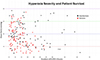 |
Figure 1 Scatter plot of patient survival by severity of hyperoxia. Each dot represents an individual patient, with survivors in red and non-survivors in black. The colored dashed lines represent the PaO2 threshold levels of 100 (red), 200 (blue), and 300 (green). |
Patient demographics and clinical characteristics stratified by hyperoxia severity.
In univariable analysis, severe hyperoxia compared to normoxia was associated with higher odds of in-hospital mortality (OR 2.7, 95% CI 1.0–7.4, p = 0.04) and the composite outcome of cardiovascular or renal complications (OR 4.3, 95% CI 0.9–20.7, p = 0.04). There was a trend towards a higher incidence of stage II or higher AKI, which did not reach statistical significance (OR 6.5, 95% CI 0.8–53.9, p = 0.06). In multivariable regression analysis adjusted for age, body surface area (BSA), and indication for VA–ECLS, severe hyperoxia remained significantly associated with the composite outcome of cardiovascular or renal complications (OR 4.6, 95% CI 0.9–23.0, p = 0.03). The association with in-hospital mortality trended towards but did not reach statistical significance (OR 2.5, 95% CI 0.9–7.4, p = 0.1) (Table 3).
Outcomes of patients undergoing VA–ECLS using a univariable and multivariable regression analysis.
Subset analysis in neonates showed similar results, with higher odds of in-hospital mortality (OR 2.7, 95% CI 0.9–8.1, p = 0.03) and cardiovascular or renal complications (OR 15.3, 95% CI 1.9–124.1, p = 0.01) in univariate analysis when comparing the severe hyperoxia group to those without hyperoxia. The association remained significant for cardiovascular or renal complications (OR 11.9, 95% CI 1.4–100.4, p = 0.03) in multivariable regression analysis. Severe hyperoxia was not associated with stage II or higher AKI, either in univariable (OR 11.3, 95% CI 0.6–221.9, p = 0.11) or multivariable analysis (OR 19.4, 95% CI 0.8–452.9, p = 0.07) (Table 4).
Outcomes of patients undergoing VA–ECLS using a univariable and multivariable regression analysis amongst neonates (age ≤ 30 days) n = 168.
New morbidity and unfavorable functional outcome for survivors who required VA–ECLS stratified by hyperoxia.
Among 126 survivors for whom functional status could be assessed, there was no statistically significant association between the severity of hyperoxia and adverse functional outcomes. The incidence of new morbidity and unfavorable outcomes was similar across all groups, p = 0.88 and p = 0.81, respectively (Table 5).
Discussion
Our study examines the outcomes of patients supported by VA–ECLS stratified by the severity of hyperoxia exposure. Nearly half of the VA–ECLS runs were performed for a cardiac indication. The overall in-hospital mortality was 45% and Stage II or higher AKI was 75.8%. Hyperoxia was more common in patients requiring VA–ECLS for a cardiac indication. Patients in the moderate hyperoxia group (PaO2 = 201–300 mmHg) were older and, consequently, had a higher weight compared to the rest of the cohort. Patients in the severe hyperoxia group had the highest in-hospital mortality rate of 64% and had the highest incidence of cardiovascular and renal complications, and stage II or higher AKI. Severe hyperoxia was associated with increased odds of composite outcome of cardiovascular or renal complications. Additionally, while severe hyperoxia was associated with higher odds of in-hospital mortality in univariable analysis, this association trended towards but did not reach statistical significance in multivariable regression analysis.
Hyperoxia potentiates the production of reactive oxygen species, which leads to lipid peroxidation, protein damage, and cell death [17]. It has been linked to adverse outcomes in various clinical scenarios. Studies have reported higher mortality and worse neurological outcomes in patients with traumatic brain injury, and infants with hypoxic ischemic encephalopathy exposed to a PaO2 level greater than 200 mmHg [1, 3]. In the critical care setting, exposure to high PaO2 levels correlates with increased mortality in both adult and pediatric populations [18–20]. Additionally, after cardiac arrest or E–CPR, hyperoxia is associated with worse outcomes, with one study demonstrating a 24% increase in mortality per 100 mmHg increase in PaO2 [4, 5, 21].
Hyperoxia during ECLS has been linked to worse outcomes. In a study of 93 infants undergoing cardiac surgery, a PaO2 of > 193 mmHg on VA ECLS within the first 48 post-operative hours was associated with higher 30-day mortality in both unadjusted (OR: 16.6, 95%CI: 3.17–305, p = 0.001) and adjusted (OR: 9.79, 95% CI: 1.18–81, p = 0.03) regression analysis. Moreover, patients exposed to hyperoxia had a higher incidence of renal replacement therapy, with an overall in-hospital mortality rate of 49% [10]. In another multi-centric study including 484 patients on VA (86.7%) and veno-venous (VV) (13.3%) ECLS, hyperoxia defined as PaO2 > 200 mmHg during the first 48 h was associated with higher mortality (OR 1.03, 95% CI: 1.01–1.04). However, no significant differences in functional status scale or renal complications were observed. Notably, patients exposed to hyperoxia were more likely to have a cardiac indication for ECLS. The in-hospital mortality was 45% in this study [9]. Similar findings have been reported in adult patients undergoing VA ECLS [22, 23]. Comparable to prior studies, the overall in-hospital mortality in our study was 45% and patients with PaO2 > 200 mmHg were older and more likely to have a cardiac indication for ECLS. This observation likely reflects the more stringent PaO2 targets typically adopted by the neonatal intensive care units compared to pediatric or cardiac intensive care units. Although severe hyperoxia was noted to be associated with in-hospital mortality in univariable analysis, this association did not remain significant in multivariable regression analysis, likely due to the small number of patients in this group. However, severe hyperoxia was significantly associated with the composite outcome of cardiovascular or renal complications during ECLS. The association between severe hyperoxia and Stage II or higher AKI trended towards significance, suggesting a potential link that requires further investigation. Consistent with the prior study by Cashen et al. [9], no difference in the functional status at discharge was noted based on hyperoxia severity.
Our study adds to the growing body of literature on the impact of early hyperoxia on outcomes in patients undergoing VA–ECLS. The first 48 h after initiating VA-ECLS are often characterized by a profound systemic inflammatory response, which can be exacerbated by hyperoxia. This early phase of ECLS is therefore a period of significant vulnerability to hyperoxia mediated organ injury. Our findings suggest a potential threshold effect with PaO2 greater than 300 mmHg, associated with adverse outcomes. The duration of exposure above this threshold may also play a role, but quantifying this clinically remains challenging. Despite technological refinements, mortality among patients supported by VA–ECLS remains high. Identifying modifiable risk factors such as hyperoxia is crucial for improving outcomes. Mitigating exposure to hyperoxia remains an important target for intervention. In the absence of robust literature, PaO2 targets vary across centers and among providers. There is significant heterogeneity in the definition of hyperoxia and the thresholds at which its deleterious effects manifest. Therefore, a collaborative effort to standardize PaO2 targets during VA–ECLS and prospectively evaluate its impact on outcomes is warranted.
Limitations
Our study is limited by its single-center retrospective design. The PaO2 targets on ECLS varied based on the provider’s discretion. Although PaO2 measurements were obtained at dedicated time intervals, it is not possible to distinguish the effect of prolonged hyperoxia exposure from those of acute spikes in PaO2 levels. Additionally, the sample size may be insufficient to fully assess the impact of hyperoxia on the outcomes. Importantly, many of these limitations can be addressed in a multicenter validation study, which our group is currently pursuing.
Conclusion
Our study demonstrates an association between hyperoxia and adverse outcomes in VA–ECLS patients, particularly among neonates and those with cardiac indication. Severe hyperoxia was associated with increased cardiovascular and renal complications. Given the variability in PaO2 targets, standardized guidelines must be formulated and investigated to validate these findings and optimize oxygen management strategies during VA ECLS.
Funding
This research received no external funding.
Conflicts of interest
The authors have no conflicts of interest and nothing to disclose.
Data availability statement
Data is not available for this article to the public per the CHOA IRB regulations.
Author contribution statement
Ashish Saini reviewed the analysis. As the lead author, he drafted the initial and revised versions of the manuscript.
Rebecca Shamah, Joshua Qian, Kasey Keane-Lerner, and Paola Rodriguez Morales performed data collection for this study.
Pranay Nayi and Adithi Shyam provided statistical expertise for analyzing and reporting the results.
Joel Davis was involved in project administration and data validation.
Mohan John provided oversight, reviewed, revised, and approved the final manuscript.
Heather Viamonte provided oversight, reviewed, revised, and approved the final manuscript.
Assad G. Beshish is the senior author of this project. He oversaw the design of the study, data collection, analysis, and drafting of the manuscript. He reviewed, revised, and approved the final manuscript.
Ethics approval
The study was approved by the CHOA Institutional Review Board (IRB# 00001239), with informed consent waived.
References
- Brenner M, Stein D, Hu P, Kufera J, Wooford M, Scalea T. Association between early hyperoxia and worse outcomes after traumatic brain injury. Arch Surg. 2012;147(11):1042–1046. [Google Scholar]
- Lilien TA, Groeneveld NS, van Etten-Jamaludin F, et al. Association of arterial hyperoxia with outcomes in critically ill children: A systematic review and meta-analysis. JAMA Netw Open. 2022;5(1):e2142105. [CrossRef] [PubMed] [Google Scholar]
- Klinger G, Beyene J, Shah P, Perlman M. Do hyperoxaemia and hypocapnia add to the risk of brain injury after intrapartum asphyxia? Arch Dis Child Fetal Neonatal Ed. 2005;90(1):F49–F52. [CrossRef] [PubMed] [Google Scholar]
- Kilgannon JH, Jones AE, Parrillo JE, et al. Relationship between supranormal oxygen tension and outcome after resuscitation from cardiac arrest. Circulation. 2011;123(23):2717–2722. [CrossRef] [PubMed] [Google Scholar]
- Elmer J, Scutella M, Pullalarevu R, et al. The association between hyperoxia and patient outcomes after cardiac arrest: Analysis of a high-resolution database. Intensive Care Med. 2015;41(1):49–57. [CrossRef] [PubMed] [Google Scholar]
- Hayes RA, Shekar K, Fraser JF. Is hyperoxaemia helping or hurting patients during extracorporeal membrane oxygenation? Review of a complex problem. Perfusion. 2013;28(3):184–193. [Google Scholar]
- Tonna JE, Boonstra PS, MacLaren G, et al. Extracorporeal Life Support Organization Registry International Report 2022: 100, 000 survivors. ASAIO J. 2024;70(2):131–143. [Google Scholar]
- Hatami S, Hefler J, Freed DH. Inflammation and oxidative stress in the context of extracorporeal cardiac and pulmonary support. Front Immunol. 2022;13:Article 831930. [Google Scholar]
- Cashen K, Reeder R, Dalton HJ, et al. Hyperoxia and hypocapnia during pediatric extracorporeal membrane oxygenation: Associations with complications, mortality, and functional status among survivors. Pediat Critical Care Med. 2018;19(3):245–253. [Google Scholar]
- Sznycer-Taub NR, Lowery R, Yu S, Owens ST, Hirsch-Romano JC, Owens GE. Hyperoxia is associated with poor outcomes in pediatric cardiac patients supported on venoarterial extracorporeal membrane oxygenation. Pediat Critical Care Med. 2016;17(4):350–358. [Google Scholar]
- Beshish AG, Jahadi O, Mello A, Yarlagadda VV, Shin AY, Kwiatkowski DM. Hyperoxia during cardiopulmonary bypass is associated with mortality in infants undergoing cardiac surgery. Pediat Critical Care Med. 2021;22(5):445–453. [Google Scholar]
- Extracorporeal Life Support Organization (ELSO) Registry Data Definitions. Improving global outcomes 2012 clinical practice guideline; 2012. Available at: https://www.elso.org/portals/0/files/new%20registry%20elso%20registry%20data%20definitions%202025.pdf Accessed September 24, 2025 [Google Scholar]
- Stevens PE, Levin A. Kidney Disease: Improving Global Outcomes Chronic Kidney Disease Guideline Development Work Group Members. Evaluation and management of chronic kidney disease: synopsis of the kidney disease: improving global outcomes 2012 clinical practice guideline. Ann Intern Med. 2013;158(11):825–830. [CrossRef] [PubMed] [Google Scholar]
- Pollack MM, Holubkov R, Glass P, et al. Functional Status Scale: new pediatric outcome measure. Pediatrics. 2009;124(1):e18–e28. [CrossRef] [PubMed] [Google Scholar]
- Beshish AG, Rodriguez Z, Farhat MH, et al. Functional status change among infants, children, and adolescents following extracorporeal life support: A multicenter report. ASAIO J. 2023;69(1):114–121. [CrossRef] [PubMed] [Google Scholar]
- Pollack MM, Holubkov R, Funai T, Clark A, Moler F, Shanley T, et al. Relationship between the functional status scale and the pediatric overall performance category and pediatric cerebral performance category scales. JAMA Pediatr. 2014;168(7):671–676. [CrossRef] [PubMed] [Google Scholar]
- Lefer DJ, Granger DN. Oxidative stress and cardiac disease. Am J Med. 2000;109(4):315–323. [Google Scholar]
- Ni YN, Wang YM, Liang BM, Liang ZA. The effect of hyperoxia on mortality in critically ill patients: A systematic review and meta analysis. BMC Pulm Med. 2019;19(1): [Google Scholar]
- Ramgopal S, Dezfulian C, Hickey RW, et al. Early hyperoxemia and outcome among critically ill children. Pediatr Crit Care Med. 2020;21(2):e129–e132. [Google Scholar]
- Ferguson LP, Durward A, Tibby SM. Relationship between arterial partial oxygen pressure after resuscitation from cardiac arrest and mortality in children. Circulation. 2012;126(3):335–342. [Google Scholar]
- Bonnemain J, Rusca M, Ltaief Z, et al. Hyperoxia during extracorporeal cardiopulmonary resuscitation for refractory cardiac arrest is associated with severe circulatory failure and increased mortality. BMC Cardiovasc Disord. 2021;21(1):542. [CrossRef] [PubMed] [Google Scholar]
- Jentzer JC, Miller PE, Alviar C, Yalamuri S, Bohman JK, Tonna JE. Exposure to arterial hyperoxia during extracorporeal membrane oxygenator support and mortality in patients with cardiogenic shock. Circ Heart Fail. 2023;16(4):Article e010328. [Google Scholar]
- Celińska-Spodar M, Załȩska-Kociȩcka M, Banaś S, et al. Arterial hyperoxia and mortality in patients undergoing venoarterial extracorporeal membrane oxygenation. Shock. 2023;59(1):20–27. [Google Scholar]
Cite this article as: Saini A, Shamah R, Qian J, Keane-Lerner K, Morales PR, Nayi P, Shyam A, Davis J, John M, Viamonte H & Beshish AG. Impact of early hyperoxia on outcomes during neonatal and pediatric veno-arterial extracorporeal life support. J Extra Corpor Technol 2026, 58, 32–38. https://doi.org/10.1051/ject/2025057.
All Tables
Patient demographics and clinical characteristics stratified by hyperoxia severity.
Outcomes of patients undergoing VA–ECLS using a univariable and multivariable regression analysis.
Outcomes of patients undergoing VA–ECLS using a univariable and multivariable regression analysis amongst neonates (age ≤ 30 days) n = 168.
New morbidity and unfavorable functional outcome for survivors who required VA–ECLS stratified by hyperoxia.
All Figures
|
Figure 1 Scatter plot of patient survival by severity of hyperoxia. Each dot represents an individual patient, with survivors in red and non-survivors in black. The colored dashed lines represent the PaO2 threshold levels of 100 (red), 200 (blue), and 300 (green). |
| In the text | |
Current usage metrics show cumulative count of Article Views (full-text article views including HTML views, PDF and ePub downloads, according to the available data) and Abstracts Views on Vision4Press platform.
Data correspond to usage on the plateform after 2015. The current usage metrics is available 48-96 hours after online publication and is updated daily on week days.
Initial download of the metrics may take a while.